

An X-ray of the neck is not one all-purpose picture. Having upper cervical X-rays explained starts with the views: a lateral image shows the neck from the side, an AP image from the front, and an open-mouth image highlights the top two neck bones. Upper cervical imaging focuses on those bones, while general cervical X-rays show the neck more broadly. Because X-rays use radiation, it is reasonable to ask why different views are taken.
Each view offers a different angle, and clinicians consider the images alongside a person’s symptoms and examination findings. Here, you’ll learn what lateral, AP, and open-mouth views can show, how clinicians interpret them together, and how upper cervical imaging differs from general chiropractic X-rays. You’ll also get a clear explanation of radiation, while keeping in view what each image can and cannot tell you.
Combining views to assess the upper cervical spine
Each cervical spine view offers a different angle on the vertebrae and their position. Clinicians interpret images alongside symptoms and examination findings, not in isolation.
Combining Multiple Views
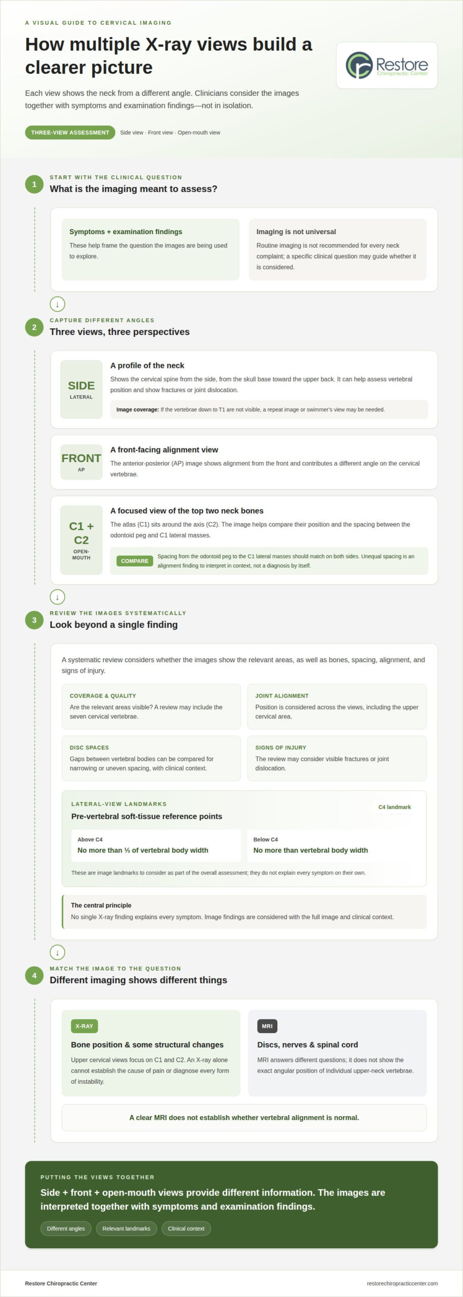
A three-view assessment commonly includes lateral, anterior-posterior (AP), and open-mouth images. The lateral view shows the neck from the side; AP and open-mouth views show alignment from the front, with the open-mouth image centered on the atlas and axis. Together, these angles provide different information about the upper cervical spine. Some clinics also use specialized projections, such as nasium or vertex views, to examine specific positioning.
An MRI answers different questions: it can show discs, nerves, and the spinal cord, but not the exact angular position of individual upper-neck vertebrae. A clear MRI therefore does not establish whether vertebral alignment is normal.
Systematic X-Ray Assessment
A systematic assessment considers image quality, bones, spacing, and signs of injury rather than focusing on a single finding. A clinician may review the seven cervical vertebrae, joint alignment, and whether the images show the relevant areas. Disc spaces are gaps between vertebral bodies; comparing them can reveal narrowing or uneven spacing that needs clinical context. No single X-ray finding explains every symptom.
How much do upper cervical X-rays cost with or without insurance? Cost depends on the imaging and coverage; the price depends on the case and is given after a quote request. The number and type of views and insurance coverage affect the amount owed.
How are they performed, and how should a patient prepare? The patient is positioned for each view and stays still while images are taken. Preparation depends on the imaging setup, so follow the instructions provided for the appointment.
When are X-rays recommended or unnecessary? Imaging may be considered when there is a specific clinical question about the neck, but routine imaging is not universally recommended for every neck complaint. Symptoms and examination findings help frame that question.
Can X-rays diagnose instability, misalignment, or neck-pain causes? X-rays can show bone position and some structural changes, but an image alone cannot establish the cause of pain or diagnose every form of instability.
Lateral, AP, and open-mouth views
A cervical spine series can show the neck from the side, front, and through an open mouth. Having upper cervical X-rays explained view by view can clarify what each image contributes.
C1 and C2 Lateral-Mass Alignment
The open-mouth view shows the atlas, or C1, around the axis, or C2, so their side portions can be compared. The distance between the odontoid peg and the C1 lateral masses should be equal on both sides. Unequal spacing is an alignment finding to interpret with the full image and clinical context, not a diagnosis by itself. This view also helps assess whether C1 or C2 sits more to one side.
Findings on the Lateral View
A lateral image shows the cervical spine in profile, allowing assessment of vertebral position from the skull base toward the upper back. In the upper cervical area, clinicians examine whether the atlas sits superior or inferior relative to the foramen magnum, and whether the axis sits posterior or inferior to it. The view can also show fractures or joint dislocation. If the vertebrae down to T1 aren’t visible, a repeat image or swimmer’s view may be needed.
Normal Measurements and Alignment Angles
A useful way to have upper cervical x-rays explained is through the landmarks each view shows. On a lateral image, pre-vertebral soft tissue above C4 should be no more than one-third of the vertebral body width. Below C4, it should be no more than the vertebral body width. On an open-mouth image, C1 lateral-mass spacing from the odontoid peg should match on both sides.
A first-visit evaluation can place these imaging details within the broader clinical picture.
Differences between upper cervical and general chiropractic X-rays
The key difference is the area and question being examined. General chiropractic X-rays may show the cervical spine broadly, while upper cervical imaging focuses on the atlas (C1) and axis (C2), the top two vertebrae. That narrower focus helps a doctor examine their position and orientation, rather than treating every neck image as interchangeable.
Image angles also matter. A standard neck X-ray taken from general angles may not show upper cervical positioning in the same way as views designed around the patient’s anatomy. For example, a focused image may help assess whether the axis sits posteriorly in relation to nearby structures. The angle changes the picture available for interpretation, not the patient’s anatomy.
In upper cervical chiropractic, measurements from these images may help determine adjustment angles and track alignment over time. Those measurements are considered alongside symptoms and examination findings, rather than treated as a stand-alone explanation for headaches, vertigo, or neck discomfort. Precision means examining the specific details an image can show while recognizing what it cannot establish.
The practical takeaway is that “chiropractic X-ray” can describe images serving different purposes. A broader cervical image surveys the neck; a focused upper cervical series examines C1 and C2 more closely. The doctor’s clinical question guides which images are relevant. Neither image type automatically explains every symptom, and an X-ray alone cannot establish the cause of a patient’s pain.
Restore Chiropractic Center: Upper cervical chiropractic
Restore Chiropractic Center provides upper cervical chiropractic care and chiropractic exams and evaluations for people seeking a clearer understanding of their spinal health. A first-visit consultation can include discussion of your symptoms and health history, along with evaluation findings, to put imaging questions into context.
If you’re considering what upper cervical X-rays may show, the next step is discussing how imaging relates to your individual evaluation. Restore Chiropractic Center also provides knee-chest upper cervical care, with care planning based on information gathered during the evaluation. This can help you understand what is being assessed and how it relates to concerns such as headaches or vertigo, without assuming an X-ray alone explains the cause.
To discuss a first-visit consultation, contact Restore Chiropractic Center.
Make your next conversation more informed
With upper cervical X-rays explained in context, the next step is connecting imaging information to the questions you want answered. You might ask what the evaluation is intended to assess, how images fit into the broader picture, and how your symptoms inform the discussion. This can make technical language easier to understand and help you take an active role in conversations about your spinal health.
If recurring headaches or vertigo are part of your experience, describing their pattern and impact can add helpful context. Restore Chiropractic Center can be a place to discuss your questions about upper cervical chiropractic and evaluation. To continue the conversation, contact Restore Chiropractic Center. Learning what an image can and cannot tell you is a constructive step toward feeling more informed about your options.
Frequently Asked Questions
What About Radiation Exposure?
Ask what clinical question the requested images are intended to answer and how each view contributes. Digital imaging keeps radiation exposure as low as reasonably achievable. This helps focus the discussion on useful information rather than taking images without a clear purpose. The number and type of images should relate to the question being evaluated.
What About Radiation From X-rays?
Remaining still helps produce a clear image, and each image takes about 2-3 seconds. The imaging process involves brief positioning for each view, rather than one continuous picture. If you’re concerned about exposure, raising that concern before imaging gives the clinician an opportunity to explain the purpose of the planned views and how they relate to the evaluation.
What Happens After X-Rays?
After imaging, the next step is understanding the observations in plain language. You can ask which structures were visible, whether the images answered the original question, and what information comes from the physical or neurological assessment instead. This separates what the pictures show from what a clinician learns through other parts of an evaluation.
What’s a Cervical Spine X-Ray?
A cervical spine X-ray is a brief imaging exam that creates pictures of the neck bones. The patient is positioned for each image and stays still during capture. The images show bone structure, while symptoms and examination findings provide additional context. That distinction helps explain why an X-ray is one part of an evaluation, not the entire assessment.
What Are Upper Cervical Spine X-Rays and What Anatomy Do They Show?
These images focus on the bony structures where the neck meets the base of the skull, including the atlas and axis. Their landmarks help describe bone position, but an X-ray does not measure nerve signals or show how the nervous system is functioning. This boundary is useful: imaging describes visible anatomy, while other assessment findings address different questions.
Disclaimer
The information in this article is provided for educational purposes only and is not intended to diagnose, treat, cure, or prevent any condition. It should not replace professional medical advice. Always seek the guidance of a qualified healthcare provider with any questions regarding your health.


